Optical magnifiers in dental medicine
Optical magnifiers in dental medicine
Good visibility of the treatment site is indispensible in all dental disciplines if high-quality results are to be achieved. Magnifying spectacles, headband magnifying instruments or a stereomicroscope should therefore be included in the instrumentarium of all treatments in which a high degree of detail precision is required.
Application
In dental treatment visibility is improved by the use of optical magnification aids: magnifying spectacles or headband magnifiers enlarge the treatment site by a factor of 2.5 to 6 times the size of the actual object, depending on the model selected. If a much higher magnification factor is required (up to 100x), the use of a stereomicroscope is recommended.
Magnifying spectacles and headband magnifiers can very easily be ergonomically positioned via spectacle frames or a headband. In the case of stereomicroscopes this is achieved using a spring-joint support, which users set according to their working posture.
Function of stereomicroscopes
With stereomicroscopes each eye observes "its image" from its own viewing angle, which is captured by a lens via the respective eyepiece. The user's brain then combines "these images" to a three-dimensional spatial picture.
Viewing the details of the restoration using magnifying spectacles, headband magnifiers or a stereomicroscope is aided by the "reflected light" of the dental lamp or cold/LED light (Light-Emitting Diode). These light sources are optionally available for magnifying spectacles and headband magnifiers and are generally integrated in stereomicroscopes. They illuminate the work area so well – assuming good quality products adjusted correctly to be shadow-free – that the user can work for a long time without suffering eye fatigue.
Options
When choosing magnifying spectacles, headband magnifiers or stereomicroscopes it is essential to note: with identical magnification (e.g. 2.5x) the viewing field expands in proportion to increases in the working distance. But: with an identical working distance the viewing field reduces. The greater the magnification – from 2.5x over 3.5x, 4x, 5x to 6x – the more the section of the working area which can be viewed shrinks.
For this reason the user should consider very carefully for which purpose the purchase will be used and if maximum magnification is necessary for each case. It can sometimes be practical to assess the magnification factor according to the area of use: a small viewing field, for example with endodontic or implantological treatment and a large viewing field with low magnification for prosthetic treatment.
Duration of observation can also be a criterion for selection. With magnifying spectacles or headband magnifiers the viewing field changes with every movement of the head – and even if it is only very minimum. With a stereomicroscope that has already been focussed, however, the viewing field remains identical over the entire duration of treatment – until the microscope is realigned. And the extra visual options of this optical magnification aid may also interest the operator, such as transmission of the viewing field to a monitor or the recording of treatment to a data carrier. The better illumination of the treatment site could also be a decisive factor for product selection in comparison with magnifying spectacles or headband magnifiers.
When purchasing magnifying spectacles, headband magnifiers or a stereomicroscope it is always recommended to try out the prospective product chairside. This allows the working distance and individual treatment position to be authentically combined and the "correct" combination of magnification factor and viewing field to be selected. This also tests the comfort of the respective product – an important criterion for daily long-term use.
Want to give it a try ...
... or need professional advice?
Get in touch with us or click Contact.
Word of the day
| English | German |
|---|---|
| center of the ridge | Kieferkammmittellinie |
Focus text of the month
Composites also composite (from the Latin componere = to compose) are tooth-coloured filling materials with plastic properties used in dental treatment. In lay terms they are often referred to as plastic fillings, also erroneously sometimes confused with ceramic… Composites also composite (from the Latin componere = to compose) are tooth-coloured filling materials with plastic properties used in dental treatment. In lay terms they are often referred to as plastic fillings, also erroneously sometimes confused with ceramic fillings due to their tooth colour. After being placed in a cavity they cure chemically or by irradiating with light or a combination of the two (dual-curing). Nowadays, composites are also used as luting materials. The working time can be regulated with light-curing systems, which is a great advantage both when placing fillings and during adhesive luting of restorations. Dual-curing luting materials are paste/paste systems with chemical and photosensitive initiators, which enable adequate curing, even in areas in which light curing is not guaranteed or controllable. Composites were manufactured in 1962 by mixing dimethacrylate (epoxy resin and methacrylic acid) with silanized quartz powder (Bowen 1963). Due to their characteristics (aesthetics and advantages of the adhesive technique) composite restorations are now used instead of amalgam fillings.
The material consists of three constituents: the resin matrix (organic component), the fillers (inorganic component) and the composite phase. The resin matrix mainly consists of Bis-GMA (bisphenol-A-glycidyldimethacrylate). As Bis-GMA is highly viscous, it is mixed in a different composition with shorter-chain monomers such as, e.g. TEGDMA (triethylene glycol dimethacrylate). The lower the proportion of Bis-GMA and the higher the proportion of TEGDMA, the higher the polymerisation shrinkage (Gonçalves et al. 2008). The use of Bis-GMA with TEGDMA increases the tensile strength but reduces the flexural strength (Asmussen & Peutzfeldt 1998). Monomers can be released from the filling material. Longer light-curing results in a better conversion rate (linking of the individual monomers) and therefore to reduced monomer release (Sideriou & Achilias 2005) The fillers are made of quartz, ceramic and/ or silicon dioxide. An increase in the amount of filler materials results in decreases in polymerisation shrinkage, coefficient of linear expansion and water absorption. In contrast, with an increase in the filler proportion there is a general rise in the compressive and tensile strengths, modulus of elasticity and wear resistance (Kim et al. 2002). The filler content in a composite is also determined by the shape of the fillers.
Composite restorations Conclusion |
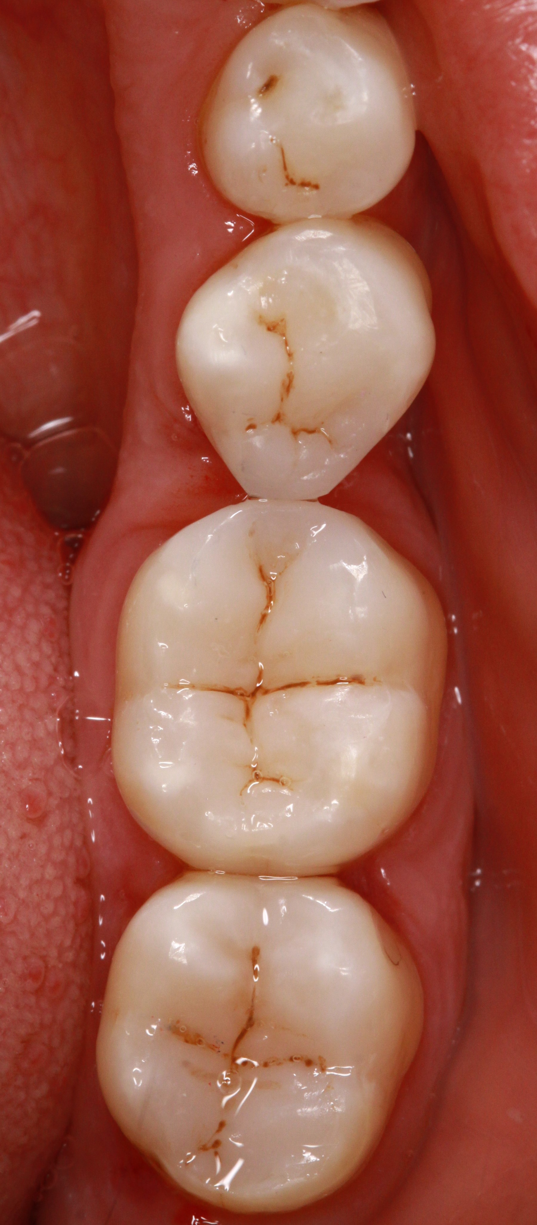
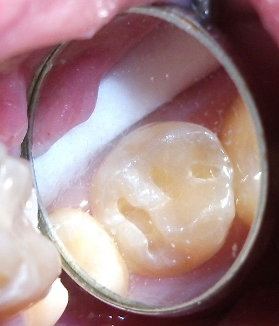 Minimally-invasive preparation and
 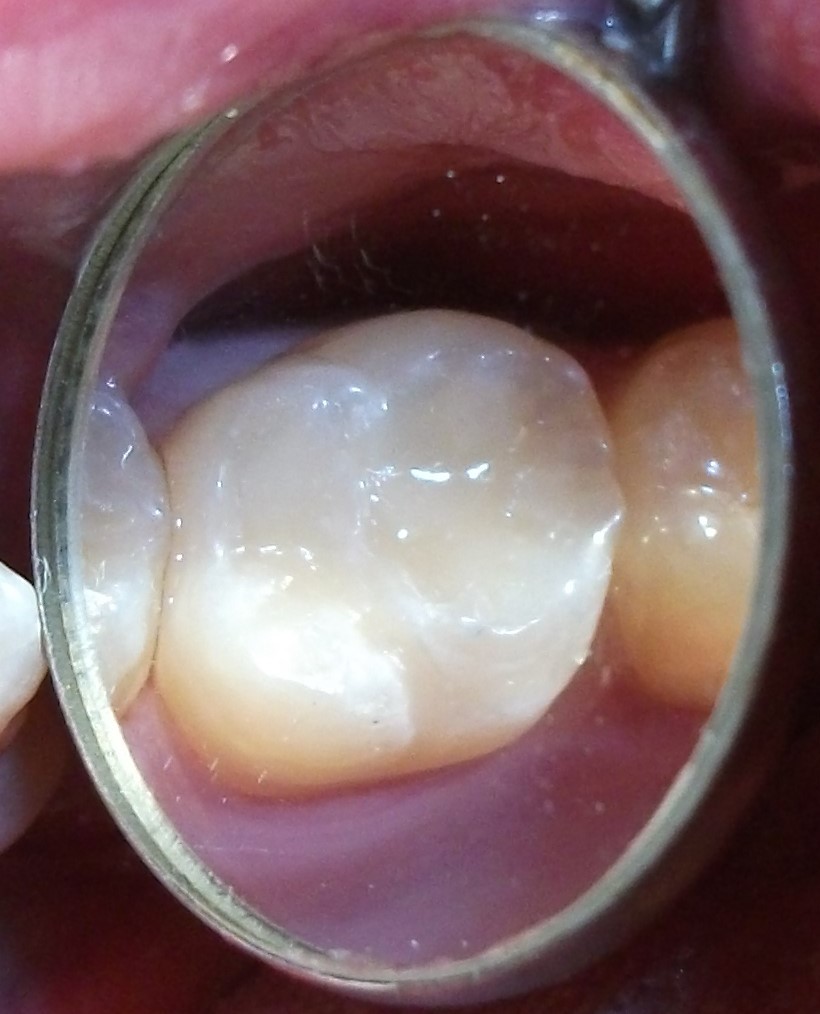 indiscernible composite restoration